RNA-seq Developmental stages gmv3.3
Gene models: v3.3
Description:
Expression data from several developmental stages for Gransden WT and Reute WT
("Dry spores" and "leaflets - Ksl" have 2 replicates and the "imbibed spores - BlqA"
only have one. All other experiments have 3 replicates).
Dry spores and imbibed spores from Grandsen and
protonema Klq, gametophore ksl and adult gametophore ksl from Reute are unpublished data.
The rest of the experiments were published in Perroud et al., 2018
| Abbreviations | |
|---|---|
| BCD liquid | Blq |
| BCDA (ammonium) liquid | BlqA |
| BCD solid | Bsl |
| BCDA (ammonium) solid | BslA |
| Gibberellin A9 methyl-ester | GA9 |
| hydroponic | hydr |
| Knop liquid | Klq |
| Knop liquid ammonium | KlqA |
| Knop solid | Ksl |
| 12-oxophytodienoic acid | OPDA |
Note: OPDA is a precursor of jasmonic acid.
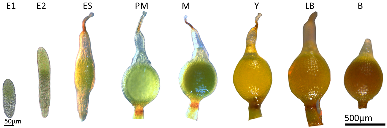
Figure 1. Sporophyte developmental stages
| Abb. | Developmental stage | Description |
| E1 | Embryo 1: |
Developing embryo; the upper, chloroplast rich half will develop into the spherical, spore-containing spore capsule, whereas the lower part will connect to the gametophore for nourishment of the developing sporophyte. |
| E2 | Embryo 2: |
Elongated embryo without developed stomata. Connection between gametophore and sporophyte still loose but cells started to differentiate. |
| ES | Early sporophyte: | Stomata are developed, clear separation of capsule (inflation) and developing seta (2n). |
| PM | Premeiotic sporophyte: | Spherical green translucend sporophyte containing spore mother cells (2n), seta starts to turn brownish. |
| M | Meiotic sporophyte: | Opaque green sporophyte containing tetrades which will later form each four spores (1n, meiosis occured). |
| Y | Yellow sporophyte: | Contains early spores of the final size covered by a plasma membrane, seta darkend during the maturation process. |
| LB | Light brown sporophyte: | Spores started to mature, the spore wall thickens by exine deposition. |
| B | Brown sporophyte: | Spores are mature, spore wall consists of a thick perine layer with spikes, exine, seperating layer and intine, sporophyte detaches easily. |
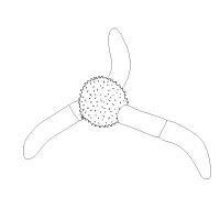
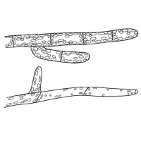
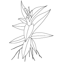
The next figures represent the available samples in this data set. The color in the colored part of the figures below will be replaced by the expression color (from the white-yellow-orange-red color scale) when using the Expression Viewer.
Gransden WT - Dry Spores

- Dry Spores
Gransden WT - Imbibed Spores BlqA
- Imbibed Spores - BlqA
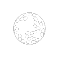
Gransden WT - Germinating Spores BslA
- Germinating spores - BslA
Gransden WT - Protonema BCD solid
- Protonema - Bsl
Gransden WT - Protonema Knop solid
- Protonema - Ksl
Gransden WT - Protonema Knop liquid
- Protonema - Klq
Reute WT - Protonema Knop liquid
- Protonema - Klq
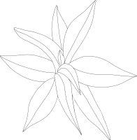
Reute WT - Gametophores Knop solid

- Gametophores - Ksl
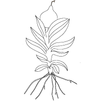
Reute WT - Adult Gametophores Knop solid
- Adult Gametophores - Ksl
Gransden WT - Gametophores no rhizoids Knop solid
- Gametophores no rhizoids - Ksl
Gransden WT - Gametophores no rhizoids BCD solid
- Gametophores no rhizoids - Bsl
Gransden WT - Leaflets Knop solid

- Leaflets - Ksl
Reute WT - Green Sporophyte Knop solid
- Green Sporophyte - Ksl
Reute WT - Brown Sporophyte Knop solid
- Brown Sporophyte - Ksl
Gransden WT - Protoplast BCDA solid
- Protoplast - BslA